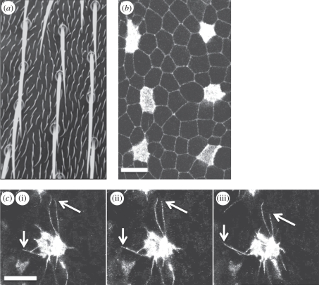
Figure 1.
Sensory organ patterning is driven by signalling between dynamic cellular protrusions. (a) A section of the notum of an adult Drosophila melanogaster fruitfly displays the evenly spaced, grid-like pattern of microchaete bristles that act as mechanosensory organs. Between each bristle are ordinary epithelial cells, each of which expresses a small hair. (b) The development of the pattern of mechanosensory organ precursor cells can be observed in fly pupae. The image shows the apical section of epithelial cells in an area of the notum close to the fly midline, at 14 h after pupae formation. Cells destined to become sensory organs (microchaete bristles) express Neuralized-Gal4, UAS-Moesin-GFP (Neu-GFP). Ubiquitously expressed E-Cadherin-GFP is used to visualize apical cell–cell junctions. (c) Confocal sections reveal the dynamic protrusions (filopodia and lamelopodia) in the basal section of a typical epithelial cell. The cell in this example is imaged through its expression of Neu-GFP. The image shows the position of filopodia at three 100 s intervals, which extend over multiple cell diameters. The arrows highlight the ends of two filopodia that can be observed extending and retracting (mean filopodia lifetime approx. 500 s—data not shown). Scale bars, 10 µm, (c) (i) 0 s, (ii) 100 s, (iii) 200 s.

