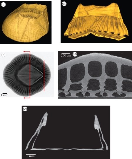
Figure 2.
X-ray radiograph and tomograms of Balanus amphitrite showing the structure of the shell. (a) Volume-rendered tomogram overview of a barnacle shell. (b) Volume renderings of a live barnacle's shell showing a quarter section from the interior with an elliptical cutout in the base to highlight the channels in the base. (c) Radiograph of a live barnacle with lines indicating the approximate regions shown in (d) and (e). (d) Tomogram two-dimensional secant slice reconstruction showing the outer lamina, longitudinal canals, longitudinal septum and inner lamina above, and the interlocking parietes and base plate structures below in a live barnacle. (e) Tomogram two-dimensional thick transverse section reconstruction, near the mid-section of a live barnacle, showing the shell cross section (the operculum has been removed for clarity). Data are from two representative live individuals and one shell. (Online version in colour.)

