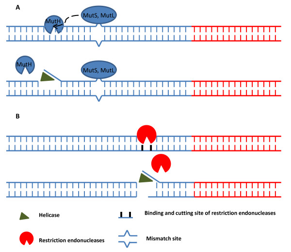
Figure 1.
Schematic model of restriction endonucleases effects on HDA. Panel A.) Mechanism in vivo: The mismatch is recognized by MutS. MutS and MutL form complex to stimulate MutH which generates nick site of the DNA duplex near the mismatch position. UvrD (helicase) is then loaded, unwinds the DNA duplex at the nick, and extends toward the mismatch. Panel B.) Mechanism in vitro: Restriction endonucleases specifically cut the DNA duplex near the target sequence, generate blunt or 5' ss or 3' ss ends or nick site (if using nicking enzyme). UvrD is then loaded and unwinds the DNA duplex. Red lines represent target DNA sequence. Blue lines represent non-target DNA sequence.

