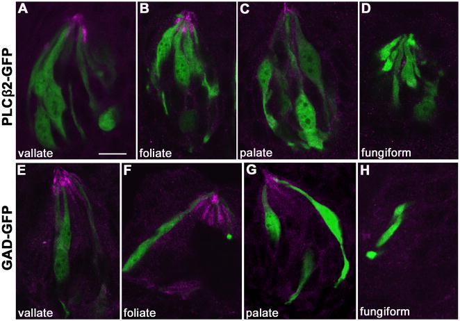
Figure 4. ROMK is not co-expressed in Type II/Receptor cells or Type III/Presynaptic cells.
A–D. Taste tissue from PLCβ2-GFP transgenic mice, was immunostained for ROMK (magenta). In confocal micrographs, immunofluorescence does not overlap with GFP-labeled Type II cells (green) in vallate (A), foliate (B), or palatal (C) taste buds. ROMK is barely detectable in fungiform taste buds (D), consistent with our qRT-PCR data (Figure 2) of minimal mRNA expression in these anterior taste buds. E–H. Taste tissue from GAD-GFP transgenic mice similarly shows a lack of ROMK immunostaining (magenta) in GFP-labeled Type III taste cells (green) in vallate (E), foliate (F) or palatal (G) taste buds. As above, ROMK was not easily detected in fungiform (H) taste buds. Only the micrograph in A was from a section with tyramide amplification (see Methods). Scale bar, 10 μm.

