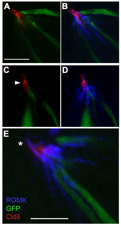
Figure 9. ROMK protein in taste cells extends to the level of apical tight junctions and beyond.
ROMK was immunodetected using anti-ROMK and Alexa 594-secondary antibody (displayed in blue pseudocolor). The taste buds are from vallate sections from a GAD-GFP mouse; GFP was immunodetected with Alexa 488-conjugated secondary antibody and is displayed green. Tight junctions were visualized using anti-Claudin-8, prelabeled with Alexa 647-Fab fragments, here displayed in red pseudocolor. Three examples of taste buds are shown with Claudin-8 and GFP overlayed (A, C), or all three antigens overlayed (B, D, E). Tight junctions (arrowhead) appear to be the apical limit for GFP, a soluble protein in the cytoplasm. In contrast, ROMK is detected in apical processes at the level of tight junctions, and occasionally extending beyond (*in E). All images are single plane (≈1μm depth) confocal micrographs. Scale bar in A applies to panels A-D; all bars, 10 μm.

