
Fig. 6.
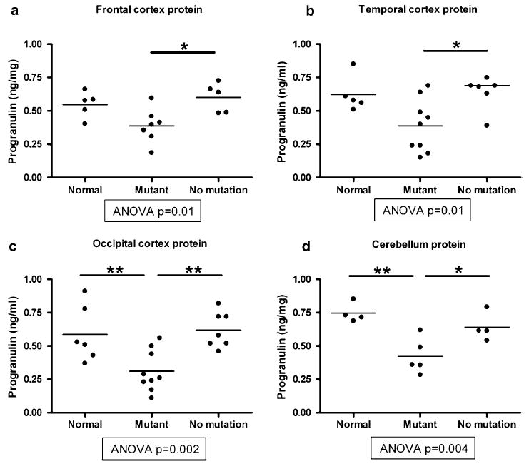
Sandwich ELISA quantification of progranulin protein levels from brain samples in normal controls (normal), FTLD-TDP with GRN mutations (mutant), and FTLD-TDP without GRN mutations (no mutation). Progranulin in human brain lysates (RIPA fraction) was captured with mAb 2161 and detected with rabbit anti-progranulin Ct antibody and HRP-conjugated goat anti-rabbit antibody. a, b In brain regions with more histopathological involvement in FTLD-TDP (frontal and temporal cortex), ELISA shows no significant difference in progranulin protein levels between normal controls and GRN mutants, although GRN mutants did differ from FTLD-TDP without GRN mutations (see bars with * comparing FTLD-TDP cases ± GRN mutations). c, d In brain regions that are relatively histopathologically spared in FTLD-TDP (occipital cortex and cerebellum), ELISA shows significantly decreased progranulin protein levels in GRN mutants relative to both normal controls and FTLD-TDP without GRN mutations (see bars with * or ** comparing GRN mutants to normal controls or FTLD-TDP cases without GRN mutations). Individual cases are shown as circles, and the mean for each group is shown as a bar. ANOVA was used to evaluate progranulin protein levels across groups, followed by Tukey’s test for pairwise comparisons *P < 0.05, **P < 0.01