
FIG 2 .
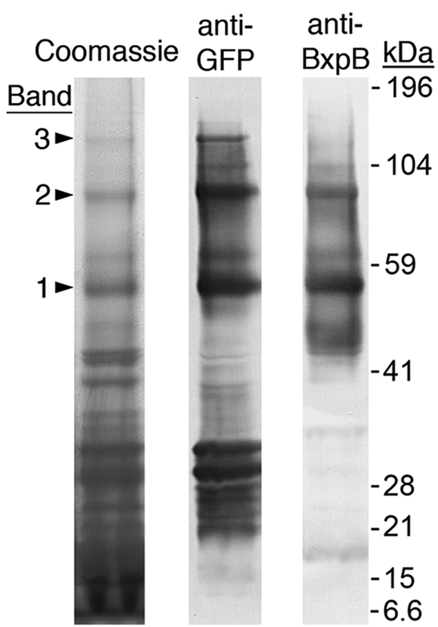
Exosporium protein complexes containing BclA NTD-eGFP fusion protein(s) attached to BxpB. After separation by SDS-PAGE, protein complexes were visualized by staining with Coomassie blue and analyzed by immunoblotting with anti-GFP and anti-BxpB MAbs. Bands 1, 2, and 3 include complexes with BxpB attached to one, two, and three molecules of the BclA NTD-eGFP fusion protein, respectively. Gel locations and molecular masses of prestained protein standards are shown. The bands in the anti-GFP lane with apparent masses of approximately 30 kDa or less presumably contain free fusion protein or products of fusion protein degradation. The bands in the anti-BxpB lane with apparent masses less than that of band 1 presumably contain BxpB complexes with other basal layer proteins or free BxpB, which has a mass of 17.3 kDa.