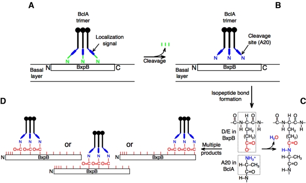
FIG 5 .
Model for the formation of isopeptide bonds that attach BclA to BxpB during exosporium assembly. (A) BclA NTD localization signals direct the binding of a BclA trimer to BxpB present in the basal layer of the exosporium. (B) Each NTD of a bound BclA trimer is proteolytically cleaved between residues S19 and A20, which produces a new and reactive amino terminus. The protein(s) required for cleavage remains to be identified. (C) The amino group of BclA residue A20 forms an isopeptide bond with an appropriately positioned side chain carboxyl group of an internal BxpB acidic residue. (D) Each strand of the BclA trimer can form an isopeptide bond with 1 of 10 acidic residues of BxpB, with each trimer presumably attaching to 3 neighboring acidic residues. There is no requirement, however, that all strands of the BclA trimer participate in isopeptide bond formation. The 13 acidic residues of BxpB are represented by red tick marks, and their positions within the protein are approximate.

