
Figure 4.
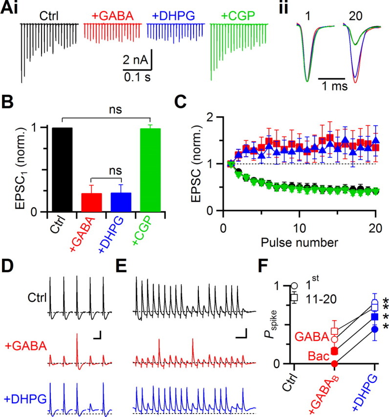
Interaction between GABABR- and mGluR-mediated modulation. A, Representative voltage-clamp experiment showing presynaptic effects of 50 μm GABA on EPSCs during 100 Hz trains of AN activation. Compared to control (black), EPSCs in GABA are reduced, and show facilitation (red). Addition of DHPG has no further effect (blue), and all the effects are blocked by the GABABR antagonist CGP55845 (green). Sample traces (i) and normalized EPSC1 and EPSC20 (ii) are shown. B, C, Average effects of GABA and DHPG from five experiments. The effects on EPSC1 amplitude (B) and normalized train EPSCs (C) are shown. The amplitude of EPSC1 does not differ significantly between GABA and GABA+DHPG (p > 0.2). The EPSC in GABA+DHPG+CGP is not significantly different from control (p > 0.3). D, E, Two representative experiments showing the interaction between mGluR and GABABR activation in current clamp. AN inputs were stimulated with single pulses (D) or a train of 20 pulses at 100 Hz (E). The effects on BC spiking were recorded in control conditions (upper traces), in the presence of GABA (middle), and in DHPG+GABA (lower). Dotted lines indicate Vrest in control conditions. Calibration: 10 mV, 20 ms. F, Average firing probabilities for experiments similar to D and E using GABA (red and blue open symbols, 6 cells), or baclofen (closed symbols, 5 cells).