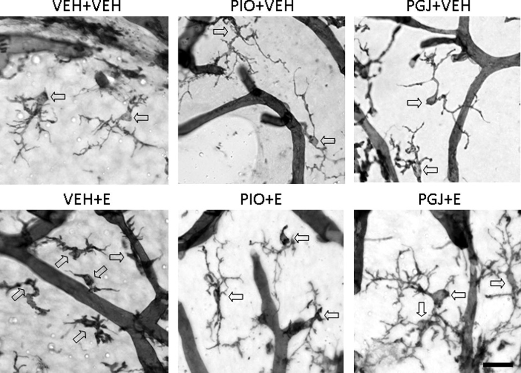
Figure 6. Morphology of microglial cells in the postnatal mouse cerebellum in vivo.
Representative micrographs of isolectin B4 stained microglial cells in the cerebellum. Animals were treated as described in Figure 5. Representative microglial cells are indicated with arrows; the isolectin B4 positive tubular structures are blood vessels. Scale bar = 25 microns.

