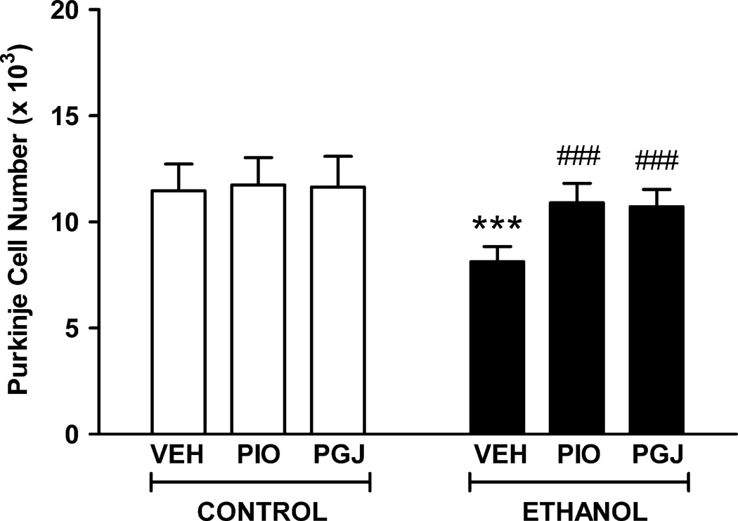
Figure 7. Protection of Purkinje cell neurons in the postnatal mouse cerebellum in vivo.
Mouse pups were administered PIO or PGJ on postnatal days 2–5. Animals were given ethanol at 3.5 g/kg on postnatal days 3–5 and tissue was harvested on postnatal day 6. Control animals were administered vehicle (VEH) in lieu of agonist or ethanol (E). Sagittal tissue sections were stained with anti-calbindin D28K antibody to identify Purkinje cells. The number of these cells in lobule IX of the cerebellar vermis was quantified using stereological methods. Results shown are the mean ± SE of six animals in each treatment group. Statistical analysis was performed with one-way ANOVA and the Fisher PLSD post hoc test. [F (5,30) = 8.932, p < 0.0001]. (*** p<0.001 compared to each of the three control conditions; ### p<0.001 compared to the VEH + ethanol condition).

