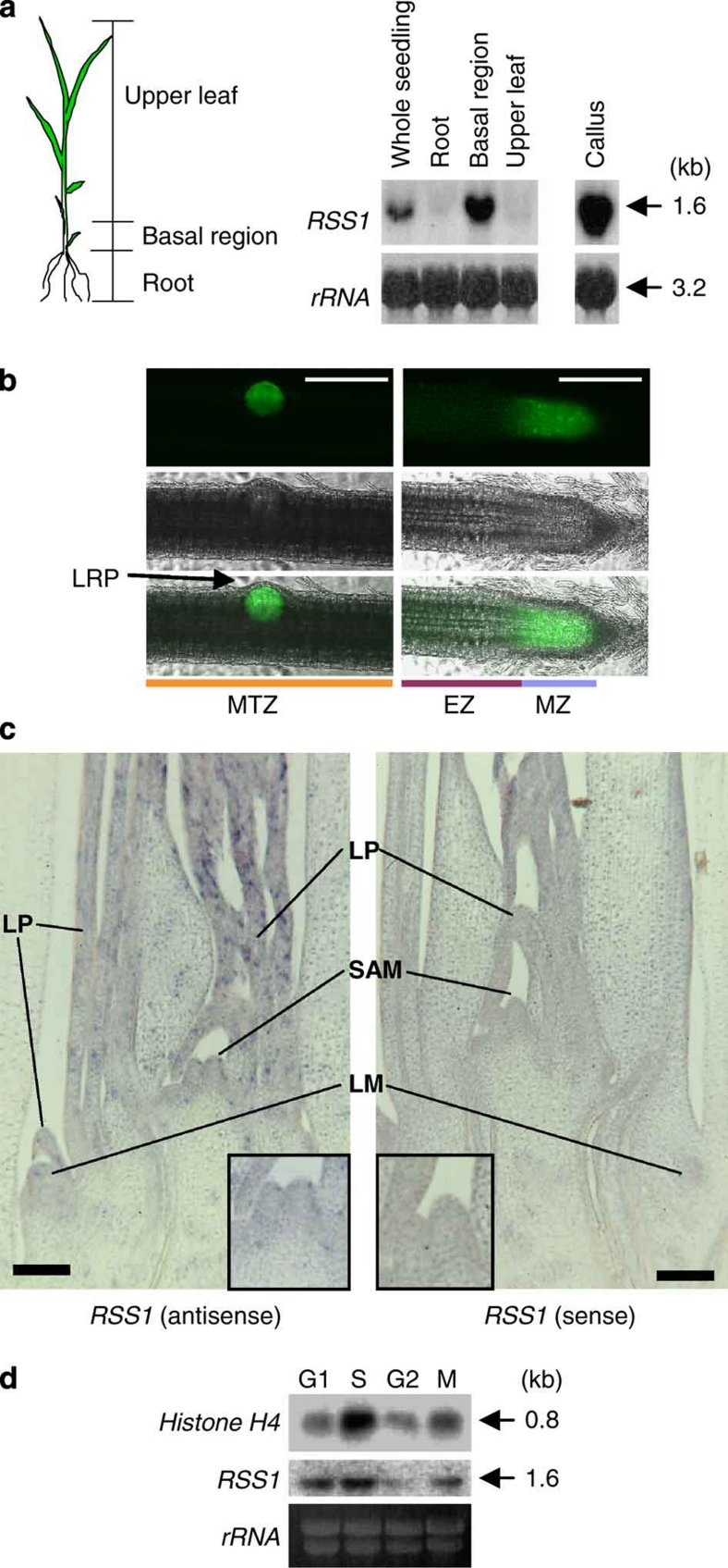
Figure 3. RSS1 is expressed preferentially in dividing cells.
(a) The expression of RSS1 in seedlings and calli. Two-week-old seedlings of rice were dissected into the three parts as in the left panel and used for northern blot analysis with an RSS1-specific probe (right). The basal region of the shoot contains the shoot apical meristem (SAM) and leaf primordia (LP). rRNA, loading control. (b) GFP signals detected in the root of transgenic rice carrying GFP driven by the RSS1 promoter. Dark-field GFP images (top) and bright-field images (middle) are merged in the panels on the bottom. (left panels) The MTZ of the root with an emerging lateral root primordium (LRP). (right panels) The apex of an adventitious root. Bars, 200 μm. (c) In situ localization of RSS1 mRNA in the shoot. Longitudinal sections were hybridized with anti-sense and sense DIG-labelled RNA probes specific for RSS1, respectively. LM, lateral meristem. Insets Increased magnification of the SAM region. Bars, 100 μm. (d) Expression of RSS1 during the cell cycle. The synchronized rice Oc cells were harvested at the indicated cell cycle phases and analysed by northern blotting using histone H4- or RSS1-specific probes. rRNA, loading control.

