Figure 4.
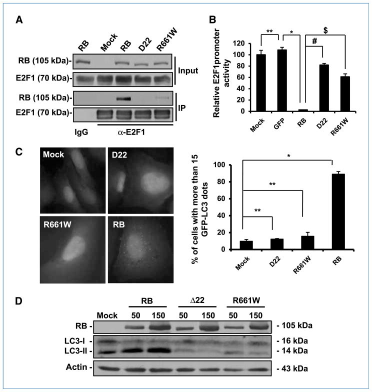
RB mutants deficient for E2F binding are unable to induce autophagy in Saos-2 Cells. A, immunoprecipitation analysis of RB and its mutants interacting with E2F1. The cells were infected with AdCMV-RB (100 pfu/cell) and AdCMV-E2F1 (5 pfu/cell), and cell lysates were collected for immunoprecipitation analysis after 48 h. B, effect of RB and RB mutants on E2F1 activity. The cells were infected with the indicated vectors (100 pfu/cell). After 24 h, the cells were transfected with a luciferase-reporting plasmid with an E2F1 promoter. Twenty-four hours later, the luciferase activity of the samples was determined (columns, mean; bars, SD). *, P = 0.0007; #, P = 0.0004; $, P = 0.003; **, P = 0.4. C, green fluorescence punctation in EGFP-LC3–expressing Saos-2 cells. The cells were infected with adenovirus vectors expressing RB and its mutants (100 pfu/cell) for 72 h. Left, representative images of the cells with the indicated treatments. Right, quantification of the cells with EGFP-LC3 dots (columns, mean; bars, SD). *, P = 0.0002; **, P > 0.05. Bar, 20 μm. D, immunoblot analysis of RB and LC3 in Saos-2 cells infected with adenoviruses expressing RB or its mutants. The cells were infected with the indicated vectors for 72 h. The numbers on the top of the protein bands represent the doses of the viruses for infection (pfu/cell). Actin was used as a loading control.

