Figure 2.
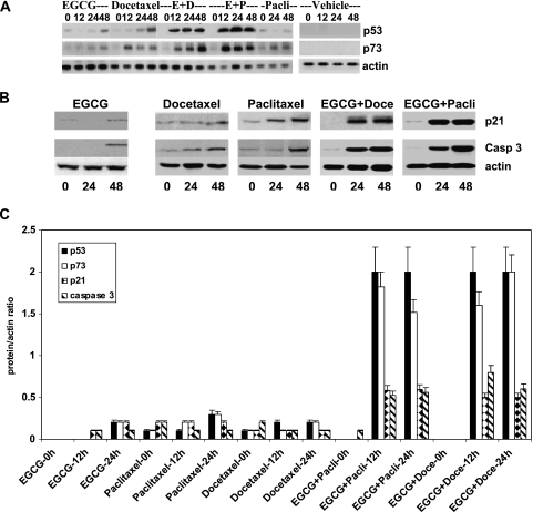
(A) Western blots of p53 and p73 expression in PC-3ML cells subjected to EGCG (30 µM), docetaxel (3.12 nM), paclitaxel (6.25 nM), and vehicle treatments alone and with (E + D) EGCG (30 µM) plus docetaxel (3.12 nM) and (E + P) EGCG (30 µM) plus paclitaxel (6.25 nM) for 0, 12, 24, and 48 hours. Control cells were treated with vehicle. (B) Western blots with p21 and caspase 3 antibodies of crude extracts from PC-3ML cells treated with EGCG (30 µM), docetaxel (3.12 nM), paclitaxel (6.25 nM), and a combination of EGCG + docetaxel or EGCG + paclitaxel at these same dosages for 0, 24, and 48 hours. (A and B) Cells were ∼70% confluent at the time of initiation of treatments. Control blots were with β-actin antibodies. (C) Calculations of the ratio of protein/actin based on densitometric scans of Western blots for p53, p73, p21, and caspase 3. PC-3ML cells at ∼70% confluence were treated with EGCG, docetaxel, paclitaxel, EGCG + paclitaxel, EGCG + docetaxel at dosages shown in B for 0, 12, and 24 hours, respectively. Data represent the mean ± SD of measurements from three independent experiments.

