
Figure 2.
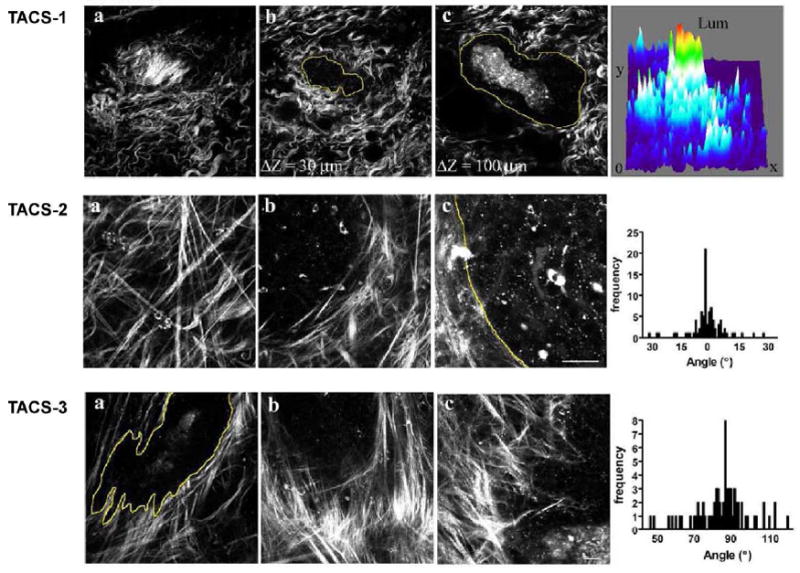
Representative methods of quantifying changes in protein fiber alignment in breast cancer by SHG microscopy. SHG probes changes in collagen in the stroma in breast cancer in a mouse model, in which disease progresses through three identifiable stages (TACS, see text) with different collagen organization. Adapted from ref. 31; images courtesy of Patricia Keely.