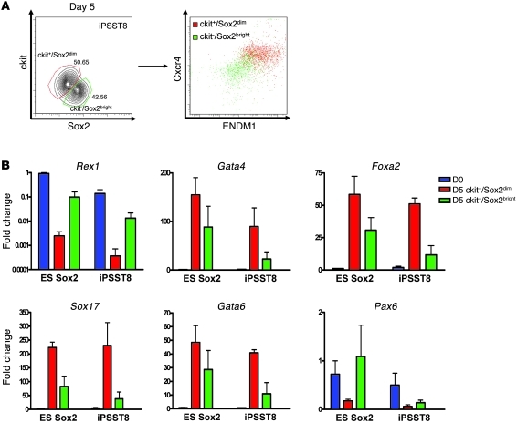
Figure 4. Methodology for purification of ES/iPS cell–derived endoderm.
(A) Representative flow cytometry analysis of ckit and Sox2 expression levels in iPS cells after 5 days of directed differentiation and expression of CXCR4 and ENDM1 cell surface markers within each indicated subgate. (B) Comparison of gene expression profiles (qRT-PCR) of Sox2-GFPdim/ckit+ and Sox2-GFPbright/ckit– sorted cell populations. Sox2-GFPdim/ckit+ fractions preferentially express endodermal gene markers, while Sox2-GFPbright/ckit– fraction expresses residual Rex1 and the neuroectodermal maker Pax6. D0, day 0 undifferentiated cells; D5, cells differentiated for 5 days. Error bars represent mean fold change in expression ± SEM.

