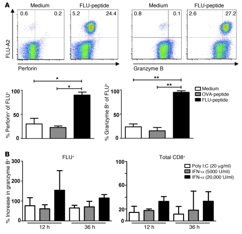
Figure 7. Lung CD8+ T cells quickly upregulate cytotoxic molecules after specific antigen contact or under influence of a type I IFN infectious milieu.
(A) LMCs were cultured for 6 days in the presence of FLU peptide, irrelevant peptide (OVA peptide), or medium. FACS plots and bar graphs show the expression of perforin and granzyme B on FLU-A2+ cells (n = 2), as measured by flow cytometry. Plots are gated on CD3+CD8+ T cells and are representative of 2 patients. Numbers indicate the percentages of CD3+CD8+ cells located in each quadrant. (B) LMCs were stimulated for 12 or 36 hours with poly I:C or different concentrations of IFN-α. Granzyme B expression was measured by flow cytometry. The percentage increase in granzyme B expression as compared with that in medium control is shown for FLU+CD3+CD8+ T cells (left) and total CD3+CD8+ T cells (right) (n = 3 patients). The error bars show SEM. *P < 0.05, **P < 0.01.

