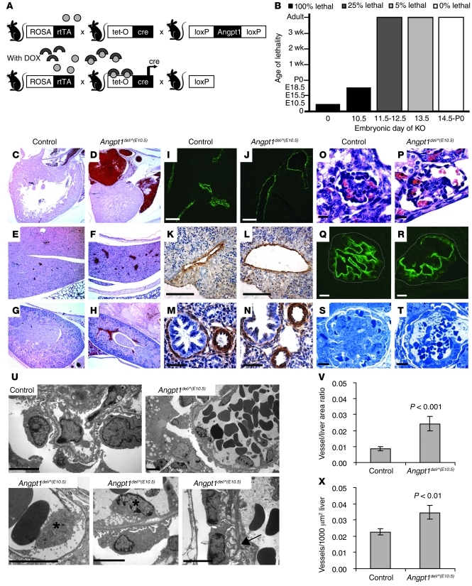
Figure 4. Angpt1 regulates both the number and diameter of developing vessels.
(A) Breeding strategy to generate inducible whole-body deletion of Angpt1 [Angpt1del/^(DOX) mice] using the DOX-inducible ROSA-rtTA/tetO-Cre bitransgenic system. (B) Induction of Angpt1 knockout at E10.5 [Angpt1del/^(E10.5) mice] or earlier results in embryonic lethality. In mice induced with DOX at E10.5, vessels are dilated at P0 (C and D) in the heart (original magnification, ×50), (E and F) liver (original magnification, ×50), and (G and H) kidney (original magnification, ×50). Pericytes/mural cells surround the vessels as shown by α-SMA staining of (I–L) liver and (M and N) lung in embryos dissected at E17.5 (See also Supplemental Figure 3). Scale bar: 10 μm (I and J); 100 μm (K and L); 50 μm (M and N). (V and X) In the same embryos, measurements of vessel number and vessel area in the liver showed a significant increase of both in Angpt1del/^(E10.5) embryos compared with those in controls. Deletion of Angpt1 at E10.5 also resulted in dilated glomerular capillary loops by E17.5, as shown by (O and P) H&E and (Q and R) podocin staining (scale bar: 10 μm), and a few glomeruli with only 1 big open capillary loop by P0, as shown by (S and T) Toluidine Blue staining (scale bar: 10 μm). (U) Electron micrographs (scale bar: 5 μm) show a folded GBM (arrow) and detachment of endothelial cells (*).

