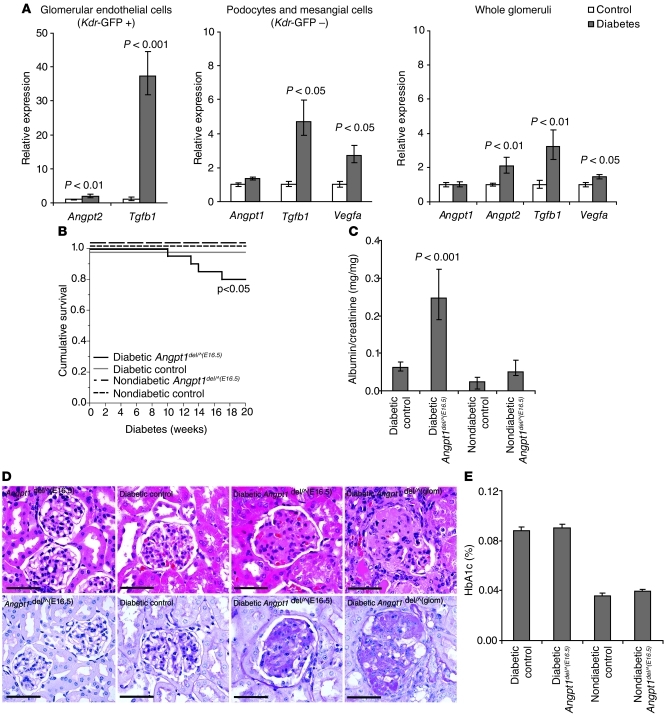
Figure 6. Angpt1 protects the glomerular vasculature in diabetic nephropathy.
(A) Expression of Angpt1, Angpt2, Tgfb1, and Vegfa in whole glomeruli or cell fractions sorted by FACS from diabetic and nondiabetic mice carrying a Kdr-GFP transgene reporter (mean ± SEM). FACS cells from glomeruli are endothelial cells in the GFP-positive fraction (Kdr-GFP +) and mainly podocytes and mesangial cells in the GFP-negative fraction (Kdr-GFP –). (B) Angpt1del/^(E16.5) mice (induced between E16.5 and P0) made diabetic show a significant decrease in survival. (C) After 20 weeks of diabetes, Angpt1del/^(E16.5) mice have a significantly higher urinary albumin/creatinine ratio compared with that of controls and nondiabetic groups. (D) Histology shows an increase in mesangial matrix expansion and sclerosis in diabetic Angpt1del/^(E16.5) mice and diabetic Angpt1del/^(glom) mice compared with that of diabetic controls (H&E, top panel; PAS bottom panel) (scale bar: 50 μm). (E) HbA1C in controls and Angpt1del/^(E16.5) mice is comparable in nondiabetic mice and after 20 weeks of diabetes.

