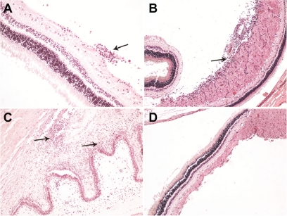
Figure 5.
Light micrographs of 4μm thick sections stained with hematoxylin-eosin revealed chronic inflammatory infiltrations (A) especially in the nerve fiber layer of the retina around the optic disc (B) and in iris (C) in a few specimens. No other histological differences were evident between injected and noninjected eyes in the rest of the specimens (D).

