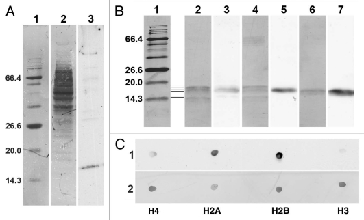
Figure 10.
Immunoblot and immunodot analysis of the reactivity of PL2-6. (A) 4–20% gradient SDS-PAGE immunoblot analysis of U2OS total cell extract. Lanes: 1, BioLab protein molecular weight (mol wt) standards, stained with Coomassie Blue (CB), indicating the mol wt (kDa) of several proteins; 2, total cell extract stained with CB; 3, ECL reaction with PL2-6. (B) 17.5% SDS-PAGE with the following lanes: 1, protein mol wt markers stained with CB; 2 and 3, HeLa core mononucleosomes; 4 and 5, equimolar mixture of recombinant Xenopus inner histones H4, H2A, H2B and H3; 6 and 7, equimolar mixture of recombinant Xenopus inner histones H2A and H2B. All lanes are from the same gel. Lanes 1, 2, 4 and 6, CB stained. Lanes 3, 5 and 7, ECL exposures carefully aligned to lanes 2, 4 and 6, respectively. Mol wt values (kDa) of the markers are indicated to the left of lane 1. The four thin horizontal lines between lanes 1 and 2 denote the positions of the four inner histones, starting with the lowest band (H4) and progressing upward, H4, H2A, H2B and H3. (C) Immunodot blots of equimolar aliquots of purified individual recombinant Xenopus inner histones (H4, H2A, H2B and H3). Strip 1, ECL reaction with PL2-6. Strip 2, identical membrane strip after CB staining.

