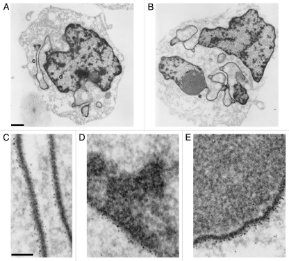
Figure 7.
Immunoelectron microscopic labeling of the epichromatin epitope at the NE periphery and within ELCS of RA treated HL-60/S4 cells. (A and B) display two different cells which exhibit nuclear lobulation and extensive formation of ELCS. Enlarged regions taken from (A and B) are as follows: (C) taken from (A) (region “c”), presents two parallel ELCS; (D) taken from (A) (region “d”), displays a segment of the nuclear surface; (E) taken from (B) (region “e”), shows a single peripheral heterochromatin layer adjacent to a nucleolus. The NE and ELCS membranes can not be visualized because of the post-fixation detergent extraction and because the samples were not fixed with OsO4. Magnification bar values: (A and B) 1 µm; (C–E) 100 nm.

