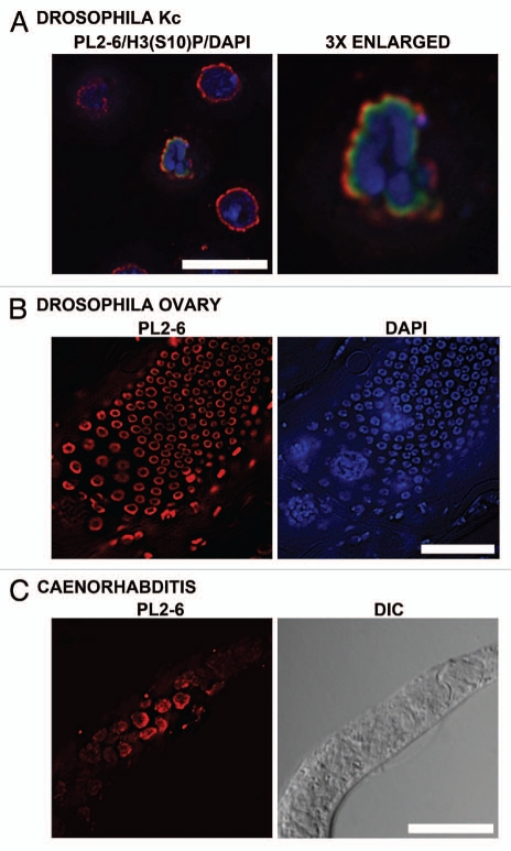
Figure 8.
Immunostaining of the epichromatin epitope in Drosophila melanogaster and C. elegans cells. (A) Drosophila Kc cells immunostained with mAb PL2-6 (red), rabbit anti-H3 phosphorylated at serine10, the mitotic marker H3(S10)p (green) and DAPI (blue). (A) (right) is a 3-fold enlargement of the mitotic chromosomes in (A) (left). (B) (left) displays PL2-6 staining (red) of Drosophila ovary cells; DAPI (blue) of the same field is shown at (B) (right). (C): C. elegans worm: left, immunostaining with PL2-6; right, DIC image. Magnification bars equal 10 µm in (A and C); 5 µm, (B).

