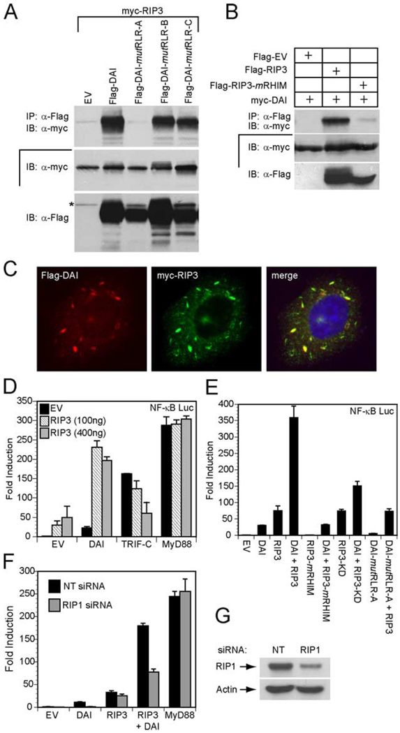
FIGURE 5.
DAI RHIM-dependent interaction with RIP3. A, Autoradiograph following IP and IB to detect mutant DAI interaction with RIP3. 293T cells were transfected with myc epitope-tagged RIP3 and Flag epitope-tagged DAI, DAI-mutRLR-A, DAI-mutRLR-B, DAI-mutRLR-C, or an EV. IP was with anti-Flag M2 beads and IB was with anti-myc Ab. IB analysis of total cell lysate confirmed the expression of myc-RIP3 and Flag-tagged proteins. The asterisk denotes residual signal of myc-RIP3. B, Autoradiograph following IP and IB to detect interaction of DAI and RIP3. 293T cells were transfected with myc epitope-tagged DAI and Flag epitope-tagged RIP3, RIP3-mRHIM, or an EV. IP used anti-Flag Ab and IB used anti-myc Ab. IB analysis of total cell lysate showed the relative expression of epitope-tagged proteins. C, Immunofluorescent localization of Flag-DAI with Myc-RIP3. Transfected HeLa cells were stained with rabbit anti-Flag Ab and with monoclonal 9E10 anti-myc Ab. Flag-DAI (red) was detected by an anti-rabbit Alexa-594-conjugated secondary Ab and Myc-RIP3 (green) was detected by an anti-mouse Alexa-488-conjugated secondary Ab. Stained cells were examined by epifluorescent microscopy (×1000). D, 293T cells were transfected with 100 ng of the pNF-κB luciferase reporter plasmid and 100 ng of Flag-DAI, Flag-TRIF, or Flag-MyD88 together with the indicated amount of Flag-RIP3 and 35 ng of the Renilla luciferase expression vectors. E, 293T cells were transfected with pNF-κB firefly luciferase reporter plasmid and 100 ng of the indicated Flag-DAI construct and/or Flag-RIP3 construct phRL-TK Renilla luciferase expression vector. F, RIP1 siRNA suppresses DAI-induced NF-κB activation. Nontargeting (NT) siRNA or RIP1 siRNA was transfected into 293T cells. At 72 h, cells were transfected with 100 ng of the pNF-κB luciferase reporter plasmid and 100 ng of Flag-DAI and/or Flag-RIP3, or Flag-MyD88 expression vector together with 5 ng of the Renilla luciferase expression vector. D–F, total DNA was held constant by adding EV. Luciferase activity assayed as described in the legend to Fig. 1. G, IB for RIP1 expression is shown using 293T cell extracts 72 h following transfection with the indicated siRNA.

