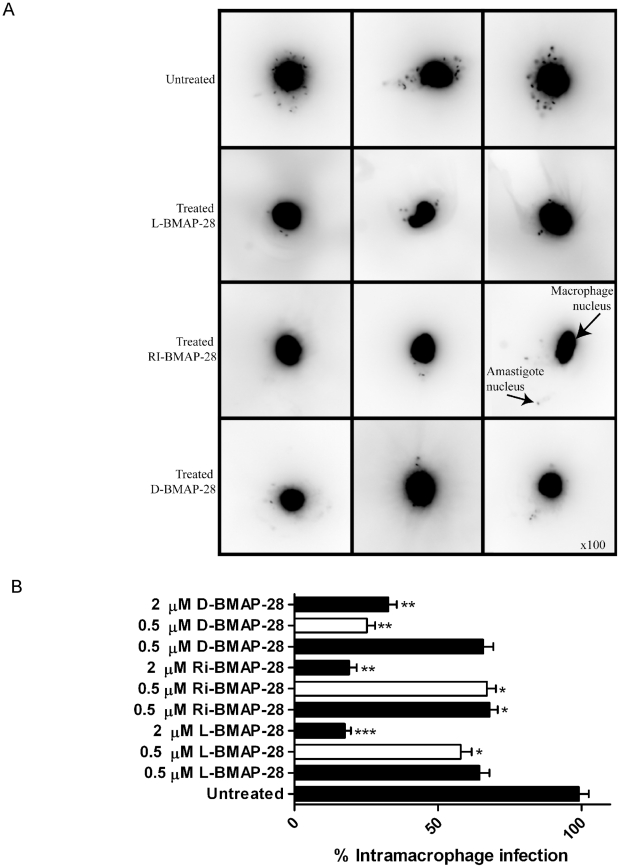
Figure 6. The effect of BMAP-28 isomers on intramacrophage L. major infection.
(A) The figure displays images that were collected from peritoneal macrophages infected with L. major Seidman wt strains for 24 h, and treated with L-, RI- and D-BMAP-28 at 0.5 µM for an additional 24 h. Infected macrophages were stained with DAPI and examined under UV light with an upright fluorescent microscope, using 100 magnification. (B) Peritoneal macrophages were infection with L. major Seidman wt (filled) and ko strains (open) for 24 h, and treated with L-, RI- and D-BMAP-28 at 0.5 and/or 2 µM for 48 h. Infections were stained with DAPI and parasite burden was quantified as an average of 100 macrophages, and expressed as a percentage of the control infection. The average number of three complete biological replicates as well as the standard errors are shown. Paired T-tests untreated vs treated indicated significance, where * p<0.05, **p<0.005, ***p<0.0005.

