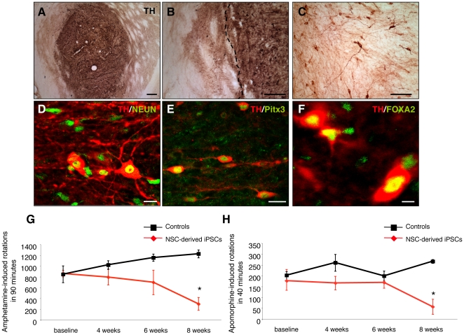
Figure 7. DA neurons from Oct4 reprogrammed adult NSCs integrate into the striatum of 6-OHDA lesioned parkinsonian rats and improve behavioural deficits.
(A) A low-power overview of an iPSC cell graft 8 weeks after transplantation stained with an antibody against TH (dark brown). (B–C) Higher magnification of a NSC-derived iPSC graft showing TH-positive soma and the reinnervation of the surrounding host striatum by donor-derived neurites. The dashed line indicates the edge of the graft. (D–F) Confocal analysis of NSC-derived iPSC grafts, 8 weeks post-transplantation, showed that most grafts contained midbrain DA neurons. The grafted TH-positive cells (red) were colabeled with antibodies against NeuN (green) (D), Pitx3 (green) (E) and FOXA2 (green) (F). (G–H) DA neurons from NSC-derived iPSCs reversed amphetamine- and apomorphine-induced rotational behaviour upon engraftment into 6-OHDA-lesioned rats. Animals were analysed for amphetamine- and apomorphine-induced rotational behaviour before and 4, 6, and 8 weeks post transplantation. Graphs show mean values±SEM. (* p≤0.05, Two-way ANOVA with post hoc analysis by Bonferroni test). Scale bars: 200 µm (A–B); 100 µm (C); 20 µm (D–E); 10 µm (F).

