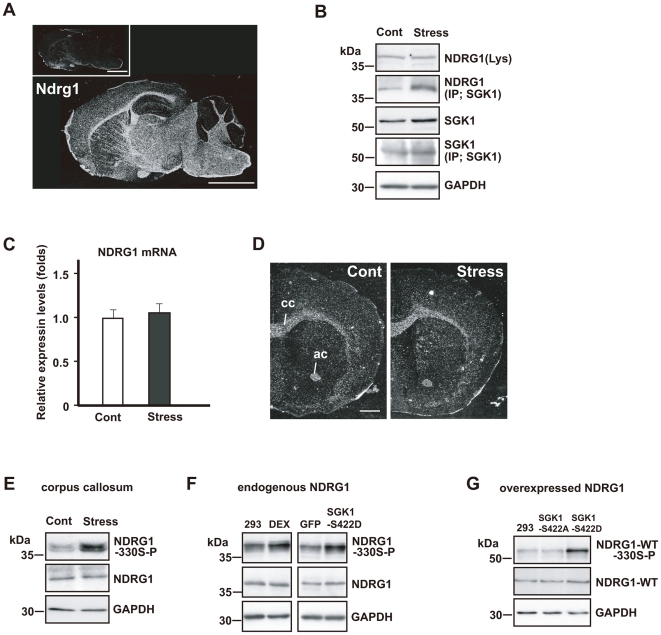
Figure 5. Repeated exposure to WIRS upregulates NDRG1 phosphorylation in the fiber tracts via SGK1 activation.
(A) In situ hybridization images of Ndrg1 mRNA. Dark-field photomicrographs show the distribution of Ndrg1 mRNA-expressing cells in the mouse brain on the sagittal plane. Sections were hybridized with 35S-labeled antisense RNA probe for Ndrg1 mRNA. As controls, adjacent sections were hybridized with 35S-labeled sense RNA probe (inset). Scale bar = 5 mm. (B) Immunoprecipitation and western blot analysis show that repeated exposure to WIRS elevated the interaction between SGK1 and NDRG1 (second column). However, NDRG1 expression did not increase in the corpus callosum (first column). (C, D) Real-time PCR analysis (C) and in situ hybridization histochemistry (D) show no alterations in Ndrg1 mRNA levels in the corpus callosum of mice exposed to repeated WIRS (see also first panel of B). cc, corpus callosum; ac, anterior commissure. Scale bar = 2 mm. (E) Western blot analysis shows that repeated exposure to WIRS elevated phosphorylated NDRG1 levels in the corpus callosum. (F) Western blot analysis shows that DEX stimulation for 24 h elevated phosphorylated NDRG1 levels in HEK293 cells (left panels). The active form of SGK1 (SGK1-S422D) or control vector (GFP) were overexpressed in HEK293 cells for 3 days (right panels). Western blot analysis shows that SGK1-S422D overexpressed cells elevated phosphorylated NDRG1 levels. 293; no-stimulation control (HEK293 cells), DEX; 100 µM DEX for 24 h. (G) The negative form of SGK1 (SGK1-S422A) or the active form of SGK1 (SGK1-S422D) were overexpressed in the HEK293 cells expressing wild type NDRG1 (NDRG1-WT) for 3 days. Western blot analysis shows that the active form of SGK1 upregulates the phosphorylation level of the overexpressed wild type NDRG1.

