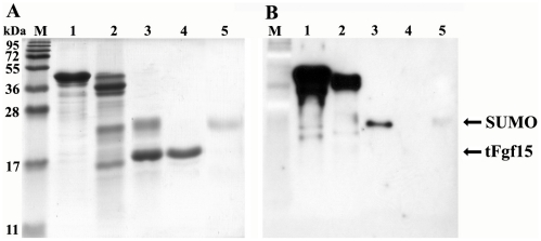
Figure 5. SUMOtFgf15 cleavage and tFgf15 purification by Ni-NTA resin.
The samples were separated on 15% SDS-PAGE gel, and stained with Coomassie Brilliant Blue (A) or undergone western blot analysis (B) with anti-His6 tag antibody. M: protein molecular weight marker, lane 1: purified SUMOtFgf15 inclusion bodies, Lane 2: refolded SUMOtFgf15, lane 3: SUMOtFgf15 digested by ScUlp1, lane 4: purified tFgf15 flow through Ni-NTA column, lane 5: eluate from Ni-NTA column using 200 mM imidazle.

