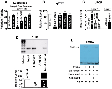
Figure 1. E2F1 as a transactivator of h-eag1 in SHSY5Y human neuroblastoma cells.
(A) Role of E2F1 in driving the h-eag1 core promoter activity. pGL3-Base: h-eag1 promoter-free pGL3 vector for control; pGL3-Core: pGL3 vector carrying the h-eag1 core promoter (a fragment spanning -630/+114); E2F1-dODN, SP1-dODN, and AP2-dODN: the decoy oligodeoxynucleotides targeting E2F1, SP1, and AP2 transcription factors, respectively, co-transfected with pGL3-Core; pGL3-Mutant: pGL3 vector carrying a mutated h-eag1 core promoter. Transfection was carried out using lipofectamine 2000. *p<0.05 vs pGL3-Core; n = 5 for each group. (B) Changes of h-eag1 mRNA level determined by real-time quantitative RT-PCR (qPCR) in SHSY5Y cells. E2F1-dODN, E2F1-MT dODN, SP1-dODN, or AP2-dODN was transfected alone. Ctl/Lipo: cells mock-treated with lipofectamine 2000; E2F1-MT dODN: the decoy oligodeoxynucleotides targeting E2F1 with mutation at the core region. *p<0.05 vs Ctl/Lipo; n = 5 for each group. (C) Increase in h-eag1 mRNA level by overexpression of E2F1 in SHSY5Y cells transfected with the plasmid expressing the E2F1 gene. E2F1-P: pRcCMV-E2F1 expression vector (Invitrogen), the plasmid carrying the E2F1 cDNA. *p<0.05 vs Ctl/Lipo; n = 5 for each group. (D) Chromatin immunoprecipitation assay (ChIP) assay for the presence of E2F1 on its cis-acting elements in the h-eag1 promoter region in SHSY5Y cells. Left panel: the bands of PCR products of the 5′-flanking region encompassing E2F1 binding sites following immunoprecipitation with the anti-E2F1 antibody or the anti-lamin A antibody for a negative control. Right panel: averaged data on the recovered DNA by anti-E2F1 expressed as fold changes over anti-lamin A band. Input: the input representing genomic DNA prior to immunoprecipitation. (E) Electrophoresis mobility shift assay (EMSA) for the fragment encompassing the putative E2F1 cis-acting element in the h-eag1 promoter region to bind E2F1 protein in the nuclear extract from SHSY5Y cells. Probe: digoxigenin (DIG)-labeled oligonucleotides fragment containing E2F1 binding site; MT Probe: DIG-labeled fragment containing mutated E2F1 site at the core motif; NE: nuclear extract from SHSY5Y cells. Solid arrowhead points to the shifted band representing the DNA-protein complex. Note that the shifted band is weakened by anti-E2F1 antibody or with the mutant E2F1 binding motif.

