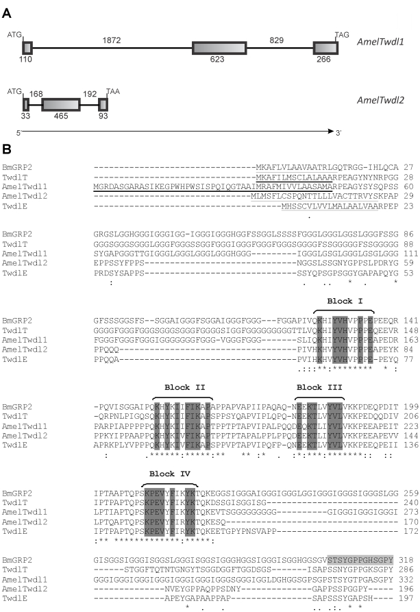
Figure 1. Tweedle: gene structures and alignment of insect tweedle proteins.
(A) Schematic representation of the AmelTwdl1 and AmelTwdl2 genes. Initiation and termination codons are indicated at the left and right of the figure, respectively. Exons and introns are indicated by boxes and lines, respectively, and the number of nucleotides is shown. The direction of transcription is indicated by an arrow. (B) Alignment (ClustalW 2) of AmelTwdl1 (ACJ38118.1) and AmelTwdl2 (ADK73965.2) amino acid sequences with other Tweedle protein sequences from B. mori, BmGRP2 (BAE06189.1), and D. melanogaster, TwdlT (AAF56656.1) and TwdlE (AAF52571.2). The four conserved blocks of amino acids are marked in dark gray. The signal peptide region is underlined in all sequences. In light gray is a region of BmGRP2 sequence that was used by Zhong et al. (see ref. [40]) to synthesize a peptide for antibody production. This antibody recognized the AmelTwdl1 protein (see Fig. 3 and corresponding text in Results section). Asterisks, colons and dots represent identical amino acid residues, strong- and weak-conservative substitutions, respectively.

