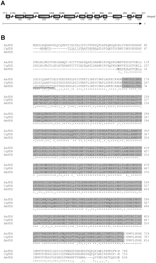
Figure 2. Peroxidase: gene structure and alignment of insect peroxidase proteins.
(A) Schematic representation of the peroxidase gene from A. mellifera. Initiation and termination codons are indicated, as well as exons (boxes) and introns (lines). The number of nucleotides is shown, and the direction of transcription is indicated by an arrow. (B) Alignment (ClustalW 2) of peroxidase sequences from A. mellifera (AmPXD, ADE45321.2), Culex quinquefasciatus (CqPXD, EDS26535.1) and Aedes aegypti (AaPXD, EAT46477.1). The signal peptide region was underlined in the A. mellifera and C. quinquefasciatus sequences. The region containing the Animal haem peroxidase domain (pfam03098) is marked in grey. Asterisks, colons and dots represent identical amino acid residues, strong- and weak-conservative substitutions, respectively.

