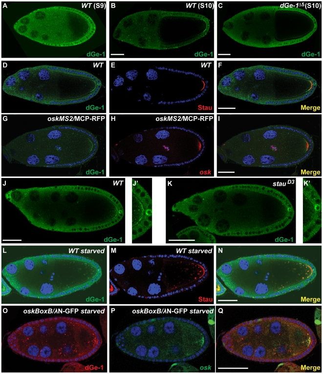
Figure 4. dGe-1 partially colocalizes with osk mRNA.
(A–C) Immunodetection of dGe-1 protein in wt (A, B) and dGe-1Δ5 GLC (C) ovaries using a rabbit anti-dGe-1 antibody. (A, B) In wt ovaries at S9 (A) or S10 (B), dGe-1 protein is enriched at the oocyte posterior pole. (C) The posterior dGe-1 signal observed in wt oocytes (B) is greatly reduced in dGe-1Δ5 GLC oocytes. (D–F) In a wt egg-chamber at late S9, dGe-1 enriched at the posterior pole of the oocyte (green, D) and partially colocalizes with Stau protein (red, E). The overlay of the two antibody signals is shown in (F). (G–I) Colocalization of dGe-1 protein (green, G) and osk mRNA (red, H) in wt S9 oocytes expressing oskMS2 mRNA and MCP-RFP protein (red, H), to which the RNA is tethered; at S9 oskMS2 mRNA distribution reflects that of endogenous osk mRNA [33]. Overlay of dGe-1 and oskMS2 mRNA signals shown in (I). (J–K) dGe-1 protein staining in wt and stauD3 oocytes. (J, J′) dGe-1 protein is enriched at the posterior pole in wt oocytes. (K and K′) In stauD3 mutant oocytes, which fail to localize osk mRNA, the amount of dGe-1 at the posterior is dramatically reduced. (L–N) Double immunostaining of dGe-1 and Stau proteins in a nutritionally restricted wt egg-chamber. In starved wt oocytes, dGe-1 protein (green, L) is present in large particles, where it colocalizes with Stau protein (red, M). Overlay of dGe-1 and Stau stainings (N). (O–Q) Distribution of dGe-1 protein and osk mRNA in starved wt egg-chambers expressing oskBoxB mRNA and λN-GFP protein, to which the RNA is tethered. In nutritionally restricted wt oocytes, dGe-1 (red, O) is present in large particles, where it partially colocalizes with oskBoxB mRNA (green, P). Overlay of dGe-1 and osk mRNA signals shown in (Q). Bar, 50 µm.

