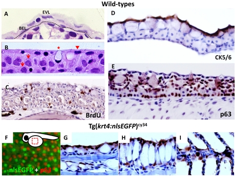
Figure 1. The krt4 promoter can target transgene expression in the superficial skin layer in zebrafish.
(A) Plastic section at 2 µm thickness showing that zebrafish skin, at 5 days post-fertilization, consists of an outer enveloping layer (EVL) and inner basal epidermal layer (BEL). The basement membrane is highlighted by the dotted line. (B) Plastic section at 2 µm thickness showing that adult zebrafish (aged at 3 months) skin consists of three major layers, including the superficial cells (arrowhead), middle and basal cells (arrows), and mucous cells (asterisk). (C) BrdU incorporation experiment showing that most skin layers in adult zebrafish are mitotically active. The BrdU+ cells (brown signals) can be detected in most skin layers at adult stage. (D) CK5/6 and (E) p63 antibodies differentially label the superficial skin layer and the putative epidermal stem cells in adult zebrafish skin, respectively. For immunohistochemistry, the 5 µm thick paraffin sections were immunostained with antigen-specific antibodies and visualized with DAB coloring substrate (brown). To visualize the cell morphology, the slides were counterstained with hematoxylin (blue). (F) Whole-mount immunostaining of p63 (red) on Tg(krt4:nlsEGFP)cy34 (green) embryonic yolk aged at 24 hpf, showing that the krt4 promoter targets the outermost EVL. The relative position of the captured image is highlighted at the upper right corner. (G–I) Immunohistochemistry of GFP (brown) on paraffin sections derived from Tg(krt4:nlsEGFP)cy34 aged at 3 months. The positive signals (brown) show that the krt4 promoter targets the superficial layer in the skin (G), esophagus (H) and gill (I) at adult stage.

