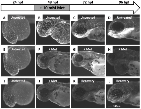
Figure 3. Administration of Met caused the killer line to lose the NTR-hKikGR+ fluorescent signals.
(A–D) The ontogenic expression of NTR-hKikGR fusion protein in killer line embryos aged from 24 hpf to 96 hpf. (E–H) Consecutive incubation of killer line embryos with 10 mM Met, from 24 hpf to 96 hpf, caused the NTR-hKikGR+ fluorescent signals to gradually diminish by 48 hpf, totally disappear by 72 hpf, and show pericardial edema in Met-treated embryos by 96 hpf. (I–L) If Met was withdrawn and replaced with fresh fish water from 48 hpf onwards, the NTR-hKikGR+ fluorescent signals partially restored by 96 hpf. Scale bar = 100 µm in L (applies to A–L). The experimental design and work flow are illustrated at the top panel. Met, metrodinazole.

