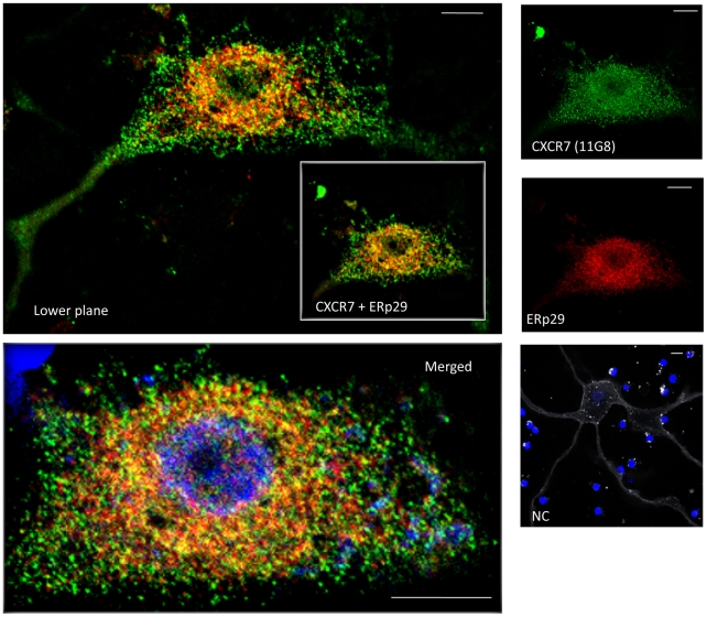
Figure 8. CXCR7 partially co-localizes with endoplasmic reticulum marker ERp29.
Human neurons were incubated with antibodies against CXCR7 (11G8, green) and ERp29 (red) as reported above and in the methods. Mouse IgG1 isotype control was used as negative control (NC). Overlapping staining of CXCR7 and ERp29 (yellow) is detected in many neurons mostly in the perinuclear region. Image labeled as “Merged” is an enlargement of the CXCR7+ERp29 image shown in the inset plus the nuclear staining, whereas the image labeled as “lower plane” is the merge of CXCR7+ERp29 from a different plane of the same cell. Scale bar: 10 µm.

