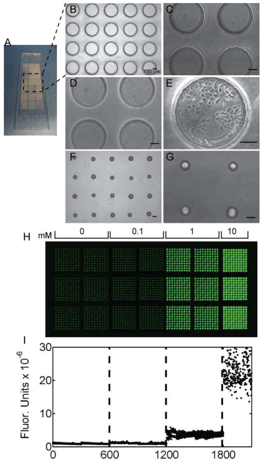
Figure 2.
Cell-seeded microwells and arrayed chemical-laden hydrogels. A) A photograph of arrayed microwells adhered to a standard glass microscope slide. B,C) Phase contrast images of microwells (400 μm in diameter and 300 μm deep) with (D,E) seeded MCF-7 breast cancer cells. F, G) Phase contrast images of arrayed PEGDA hydrogels on a PDMS substrate. All scale bars are 100 μm. H) Fluorescent scanner image of arrayed hydrogels with varying concentrations of Rhodamine B (Ex:Em, 532:575/25; green false color). The hydrogel microarray contains 600 spots of each of 0, 0.1, and 1 mM and 300 spots of 10 mM of Rhodamine B. I) Quantification of fluorescence in each arrayed hydrogel.

