
Figure 3. Photomicrographs of diencephalic regions receiving genitosensory information in a male and female rat.
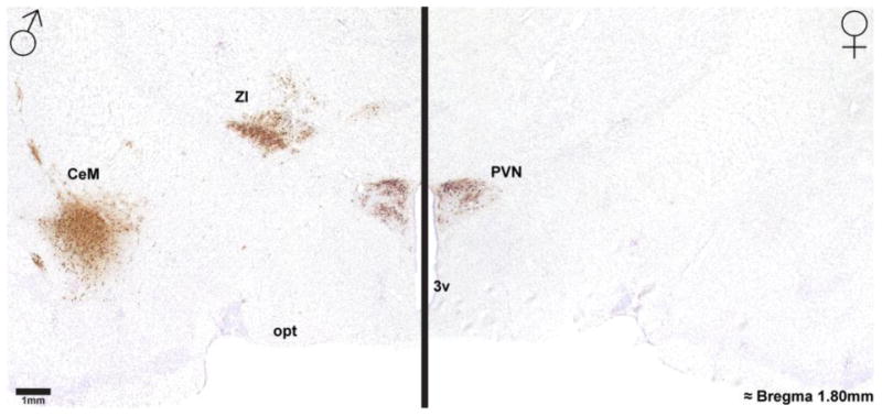
Photomicrographs of H129 labeling in the diencephalon of a male (left) and female (right) rat, as a result of genital inoculation, show robust labeling in the thalamus and central amygdala in a male but not a female rat. Note that both sexes show robust H129 labeling in the paraventricular hypothalamic nucleus (PVN). 3v = 3rd ventricle, CeM = central amygdala, opt = optic tract, ZI = zona incerta.