
Fig.3.
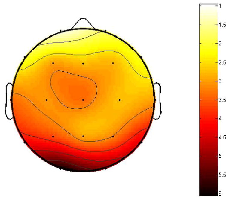
The scalp distribution of the t-scores of SEPFS difference before and after a concussive episode, note the most pronounced effect (e.g., significant decreased of SEPFS values) after concussion at electrode sites representing occipital, temporal and parietal regions.