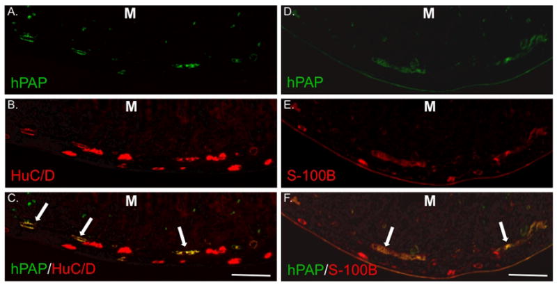
Figure 3. IP injection of isolated eNCSCs results in widespread neuronal and glial engraftment in the distal small intestine ENS.

Sections from the distal third of the small intestine 21 days after IP injection of a 7 day-old HD pup with 5 × 10⁴ hPAP+ eNCSCs. All sections are oriented with the mucosa (M) at the top of the photo. Sections were labeled immunohistochemically for hPAP (A and D), HuC/D (B), or S-100B (E). Co-labeling is illustrated in panels C and F. Long arrows indicate co-expression of markers in the region of the submucosal plexus. Bar = 200 μm.
