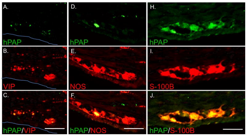
Figure 6. Engrafted neurons participate in ganglia with endogenous neurons and express VIP and nNOS while engrafted glia diffusely populate ganglia.

Views of individual myenteric ganglia in the distal small intestine of a HD rat injected with 1.5 × 10⁴ hPAP+ eNCSCs on day-of-life 2. Expression of hPAP (A, D, and H) was often noted in a small subset of cells within individual ganglia. Immunohistochemical labeling for VIP (B) and NOS (E) demonstrates the presence of both endogenous neurons and exogenous neurons within the ganglia (C and F). The glial marker S-100B most often labeled cells throughout the ganglia (I) suggesting a more clustered engraftment pattern. All sections are oriented with mucosa toward the top of the photo. The blue line indicates the serosal surface in A–C. Bar = 50 μm.
