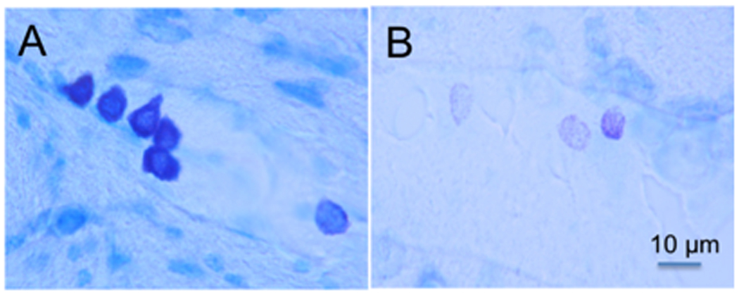
Figure 2.

Mast cells in the CNS. (A) Cluster of darkly pigmented mast cells stained with toluidine blue (purplish black) in the thalamus of a mouse. The 5 mast cells on the left are typical of the fully granulated mast cells in the mouse thalamus. The lighter cell on the right in panel A depicts a mast cell that is partially degranulated containing only scattered granules. (B) Three partially degranulated mast cells appear as stippled (pinkish-violet) cells on a background (blue) of pale parenchymal cells. Individual granules that do not touch each other are visible in these cells at higher magnification. Magnification of both photographs is indicated by the bar in panel B. (For interpretation of the references to colour in this figure legend, the reader is referred to the web version of this article.)
