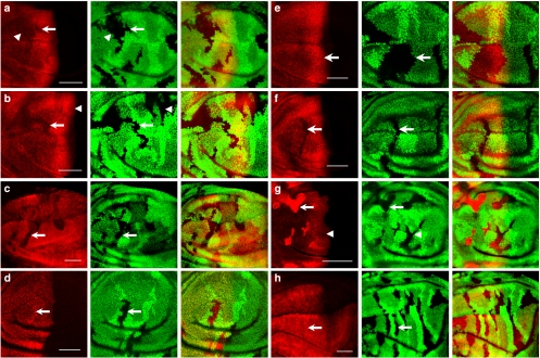
Figure 2. The CSN positively regulates Ci155 protein stability in wing discs.
(a) CSN5null clones are revealed by the absence of GFP (green) in wing discs. Ci155 stained with the 2A1 antibody (red) is reduced in CSN5null clones located in the A compartment, with arrow indicating mutant cells in the low-to-intermediate Hh signalling region and arrowhead indicating cells in the low Hh signalling region. (b) GFP-negative CSN4null clones were generated in wing discs (green). Ci155 staining (red) is lower in the CSN4null cells in the A compartment (arrow) than in the wild-type cells. The Ci155 levels in CSN4null and wild-type cells located in A/P boundary are similar (arrowhead). (c) CSN4null clones revealed by the absence of GFP (green) were generated in wing discs that simultaneously express UAS-ci-myc under the control of ms1096-GAL4. Protein levels of Ci-Myc detected by the anti-Myc antibody (red) are reduced in CSN4null mutant clones (arrow). (d) The ci-lacZ expression (red) is not reduced in CSN4null mutant clones (arrow) generated in wing discs. (e) GFP-negative CSN5null clones (green) were generated in wing discs expressing wild-type CSN5 construct under the control of ms1096-GAL4. Expression of wild-type CSN5 rescues the Ci155 level (red) in CSN5null clones (arrow). (f) Expression of CSN5D148N, which loses deneddylation activity, fails to rescues the Ci155 level (red) in CSN5null clones (arrows). (g) slimbP1493, CSN5null clones revealed by the absence of GFP (green) in wing discs show Ci155 (red) accumulation in the A compartment (arrow) except when located adjacent to the A/P boundary (arrowhead). (h) The Ci-3P mutant protein (red), carrying mutations in three PKA phosphorylation sites, is ectopically expressed in CSN4null mosaic wing pouches under the control of ms1096-GAL4. There is no difference in Ci-3P expression in wild-type and GFP-negative CSN4null cells (arrow). Scale bars in all panels represent 50 μm.

