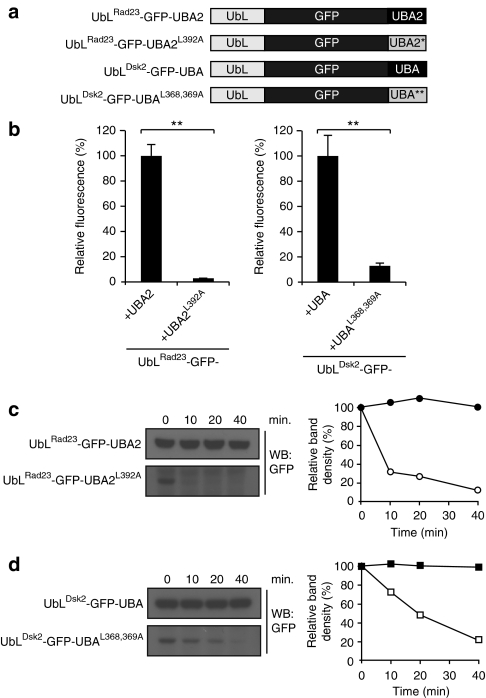
Figure 7. Protective UBA domains cannot function as initiation sites.
(a) Schematic drawing of the UbL-GFP fusions. (b) Flow cytometric quantification of the mean fluorescence intensities of yeast expressing UbLRad23-GFP-UBA2, UbLRad23-GFP-UBA2L392A, UbLDsk2-GFP-UBA or UbLDsk2-GFP-UBAL368,369A. UbLRad23-GFP-UBA2 and UbLDsk2-GFP-UBA were standardized as 100%. Values are means and standard deviations (n=3). **P<0.01 (Student's t-test). (c) Turnover of UbLRad23GFP-UBA2 (closed circles) and, UbLRad23GFP-UBA2L392A (open circles). Samples were collected at the indicated time points and detected with a GFP-specific antibody. Densitometric quantification of the blot is shown to the right. (d) Turnover of UbLDsk2-GFP-UBA (closed squares) and UbLDsk2-GFP-UBAL368,369A (open squares). Samples were collected at the indicated time points and analysed by western blotting with a GFP-specific antibody. Densitometric quantification of the blot is shown to the right.

