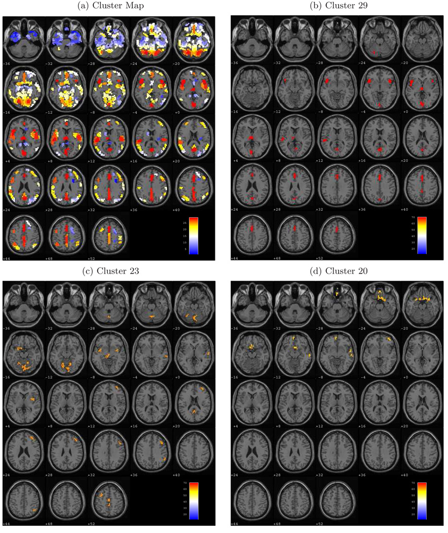
Figure 1.
Cluster maps from Ward’s method displayed on axial brain slices ranging from −36mm to +52mm relative to the anterior/posterior commissure plane (z). (a) View of the 29 clusters, with each cluster containing voxels that exhibit similar responses across varying loads on working memory. (b) Cluster 29 includes voxels in the inferior frontal gyrus (BA 47), thalamus, anterior and posterior cingulate (BA 30;32), and the lingual gyrus (BA 17;18). (c) Cluster 23 consists of voxels in the precentral and middle frontal gyri (BA 9), in the middle, medial, and superior frontal gyri (BA 6), and along the supramarginal gyrus (BA 40). (d) Cluster 20 spans portions of the right middle frontal gyrus (BA 10), the left medial frontal gyrus (BA 11), and the anterior cingulate (BA 25).

