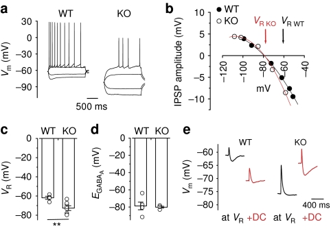
Figure 3. HCN1 deletion results in the loss of the hyperpolarizing effects of GABAA receptor-mediated inhibition.
(a) Example traces showing voltage responses to current steps injections in wild-type (WT) mice and HCN1 knockout (KO) mice. (b) An example plot showing that, unlike WT mice, HCN1 KO mice lacked a hyperpolarizing GABAA receptor-mediated driving force. Data points were fitted with a second-order polynomial function. (c) Comparison of VR in HCN1 KO mice and WT mice (WT: n=5; KO: n=8). (d) Summary plot of EGABA(A) in both genotypes (WT: n=5; KO: n=4). (e) Sample traces showing that the KO mice displayed no hyperpolarizing phase at VR, whereas WT animals had a biphasic EPSP–IPSP sequence. The hyperpolarizing IPSPs became apparent in the KO mice when cells were depolarized by DC injection. Conversely, the IPSP was less evident when pyramidal cells from WT mice were hyperpolarized. Error bars represent s.e.m.; **P<0.01.

