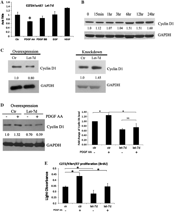
Figure 2.
PDGF-AA down-regulates let-7d expression. (A) qPCR quantifies let-7d expression in C272/hTert/E7 cells in response to PDGF-AA, PDGF-BB, VEGF and EGF. RNU49 was used as control. (B) Western blotting for cyclin-D1 in C272/hTert/E7 cells treated with PDGF-AA. (C) Western blotting for cyclin D1 in U118MG cells transfected with let-7d precursor or control (left panel). Western blotting for cyclin D1 in U118MG cells transfected with LNA targeting let-7d or scrambled LNA (right panel). (D) Western blotting assessed cyclin D1 in C272/hTERT/E7 cells treated with PDGF-AA for 6 h after transfection of let-7d precursor or control (left panel). Densitometry quantifies cyclin D1 expression levels relative to GAPDH in independent experiments. Statistical significance is indicated by asterisks (NS, not significant). (E) BRDU assay quantifies cell proliferation of C272/hTert/E7 cells transfected with let-7d precursor and control and stimulated with PDGF-AA or vehicle Statistical significance is marked by asterisks.

