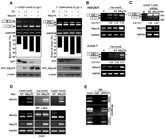
Figure 2.
NSrp70 influences alternative splice site selection of several minigenes in different cell types. (A) Promotion of exon v2 exclusion on Tra2β1 and exon v5 inclusion on CD44 minigene by NSrp70. HEK293T cells were cotransfected with increasing amounts of pEGFP/NSrp70 (0–2 µg) and 2 µg of the minigene. Parental vector (0–2 µg) was added to ensure that similar amounts of cDNAs were transfected. Exon inclusion or exclusion was determined by RT–PCR (top), as described in ‘Materials and Methods’ section. The ratio of exclusion or inclusion of Tra2β1 or CD44 minigene was shown as a histogram (middle). Dose-dependent expression of GFP_NSrp70 and GFP was confirmed by western blotting (bottom). (B) NSrp70 promotes exon v6 inclusion on Fas minigene in HEK293T or Jurkat T cells. Two micrograms of Fas minigene was transfected with 2 µg of pEGFP/NSrp70 or pEGFP into HEK293T or Jurkat T cells. Exon inclusion or exclusion was determined by RT–PCR. Note: P, parent; EV, pEGFP vector; NSrp70, pEGFP/NSrp70. Asterisks denote an uncharacterized PCR product (A and B). (C) Interference of NSrp70 by shRNA decreases exon v6 inclusion in Jurkat T cells. psiLv-H1/scramble or psiLV-H1/shNSrp70 (CCDC55) vector was infected to Jurkat T cells by the lentiviral system. Exon inclusion or exclusion was determined by RT–PCR. Note: M, 100 bp DNA marker; SR, psiLv-H1/scramble, NSrp70, psiLV-H1/shNSrp70 infection. The ratio of exon inclusion or exclusion splicing forms was analyzed by ImageJ. Each experiment was repeated at least three times to confirm the reproducibility of data. (D and E) mRNA of minigenes interacts to NSrp70. HEK293T cells were transfected with (D) or without (E) plasmid expressing myc_NSrp70 and indicated minigene and then immunoprecipitated with anti-Myc (D) or anti-NSrp70 antibody (E). The RNAs in the immunoprecipitates were then analyzed by RT–PCR using primers specific for the CD44, Fas or Tra2β1 transcripts of minigenes. Ten microliter of the supernatant of total lysate was used for total input sample.

