
Figure 5.
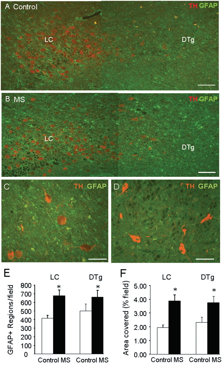
GFAP staining is increased in the locus coeruleus region of multiple sclerosis brains. Serial coronal sections through the locus coeruleus (LC) were prepared from five patients with multiple sclerosis (MS) and six controls, and stained for tyrosine hydroxylase (TH) and GFAP. The fourth ventricle is located above and to the right. Representative images from control (A) and patients (B) with multiple sclerosis show increased GFAP positive staining in the locus coeruleus and adjacent area (containing the dorsal tegmental nuclei, DTg). Representative images from one multiple sclerosis sample showing presence of GFAP staining around tyrosine hydroxylase positive stained neurons in locus coeruleus (C) but not in adjacent central pons (D). Quantitation of staining showed a significant increase in (E) the number of GFAP positive stained objects (cell bodies and processes) and (F) the total area stained (per cent field of view) in both the locus ceruleus and the dorsal tegmental nuclei of multiple sclerosis samples versus controls. Data are means ± SEM of nine sections per brain; *P < 0.05 versus controls. Scale bars are 200 µm in A and B and 100 µm in C and D.