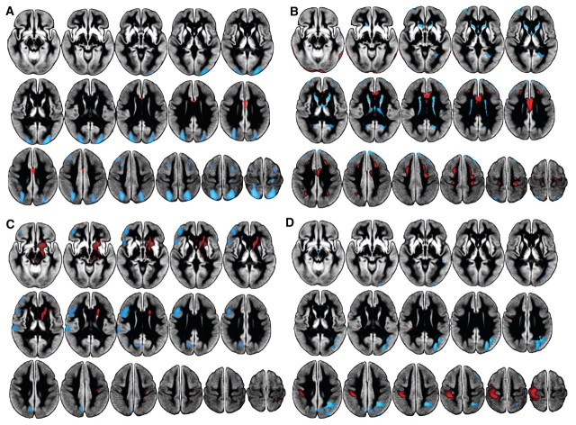
Figure 1.
Parkinson’s disease-related principal components, displayed on the study-specific average grey matter image. Black regions represent masked white matter and CSF. (A) Principal component 1 showed decreased perfusion (blue) in subjects with Parkinson’s disease relative to controls in posterior parieto-occipital regions, middle and superior frontal gyri (Broadmann areas 6 and 8), and post-central gyri, and preserved perfusion in anterior cingulate (red). Component 1 was significantly related to cognition (MoCA) and age. (B) Principal component 4 exhibited decreased perfusion primarily in the caudate and thalamus, with preferentially preserved perfusion in anterior cingulate, pre- and post-central gyri, lateral temporal and inferior occipital regions. (C) Principal component 5 showed decreased perfusion in left precuneus, cuneus, middle and inferior frontal gyri, frontal pole and operculum, and supramarginal gyri, with associated preservation in right post-central gyrus and subcortically including brainstem, caudate and putamen. Component 5 was significantly related to motor impairment (UPDRS-III). (D) Principal component 6 revealed large decreases in posterior parietal regions, precuneus, lateral occipital regions and middle frontal gyrus, with preserved perfusion in pre- and post-central gyri. Component 6 was significantly related to both motor and cognitive status. Components 2 and 3 were not related to Parkinson’s disease.

