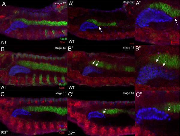
Figure 6. Migration of LVM founder cells between the CVM and salivary gland is delayed in β2t mutant embryos.
In wild-type embryos at stage 12 (A, A' and A”), LVM founder cells (LVMFs) (A' and A”, arrows) migrate anteriorly dorsal and ventral to the CVM. During stage 13 in wild-type embryos (B, B' and B”), LVMFs (B' and B”, arrows) migrate between the CVM and salivary gland. In β2tN mutant embryos at stage 13 (C, C' and C”), LVMFs have migrated anteriorly along the CVM (C' and C”, arrows) but have not migrated between the gland and CVM. Embryos in A–C were stained for Fkh (blue) to mark salivary gland nuclei, FasIII (green) for the CVM and Couch potato (red) to mark LVMFs. Scale bars in A' and A” represent 10 μm. A, B and C are projections of between 10 and 14 one micron-thick confocal optical sections whereas remaining panels depict a single one-micron thick optical section.

